

The working voltages in this microscope are 200 and 300 kV.
The expertise of our scientific and technical staff is also offered to researchers from public and private research centers and also to professionals from industrial sectors that require the use of this instrument.

Image (Resolution 0.19 nm)
- Size and morphology information (TEM).
- Crystalline Structure (Electron Diffraction and High Resolution TEM).
- Composition: Scanning-Transmission imaging with a High Angle Annular Dark Field detector (STEM-HAADF). The contrast in the image depends on the atomic number (Z-contrast). Energy Filtered TEM (EFTEM) yields information about a specific element. Composition: Scanning-Transmission imaging with a High Angle Annular Dark Field detector (STEM-HAADF).
- 3D reconstruction: electron tomography.
Chemical Analysis
- X-Ray (EDS) and Electron Energy Loss Spectroscopies (EELS).
- Combined with the STEM mode: chemical composition with spatial resolution; point spectra and composition profiles.
Vector Field analysis
- Stress and strain studies by HRTEM imaging.
In situ Physical properties measurements
- Changes of crystalline phase (Electron diffraction)
- Defect structure by Bright Field/Dark Field imaging (BF/DF) and Weak Beam imaging (WBDF).
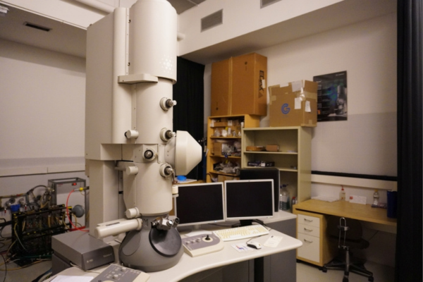
This 300 kV Field Emission Gun (FEG) TEM is fitted with a SuperTwin® lens allowing a point resolution of 1.9 Å. This TEM can work in TEM and STEM mode. For Z-contrast imaging in STEM mode, it is fitted with a High-Angle Annular Dark Field (HAADF) detector. This TEM is equipped for spectroscopy experiments performed either in EDS (X-Ray Microanalysis) or in Electron Energy Loss spectroscopy (EELS). For the latter, it is fitted with the “Tridiem” Gatan Energy Filter (GIF). This EELS set-up allows Energy Filtered TEM (EFTEM) images to be recorded as well as line spectra or spectrum imaging experiments to be performed. A 2k x 2k Ultrascan CCD camera (Gatan) is located before the GIF for TEM imaging.
In addition to these capabilities the F30 TEM is also fitted with a Lorentz lens which permit the study of magnetic materials in an environment free of magnetic field (for magnetic domain imaging). Furthermore, the F30 allows tomography experiments to be performed both in TEM and STEM mode using a dedicated single tilt holder (+/- 70°) from Fischione.

Electron tomography reconstruction from a HGNP, a rough surface caused by fast galvanic replacement is shown. Ref.: Lab On Chip 2014, 14, 325-332
doi:10.1039/C3LC50999K

Spatial distribution of the elements of immuno-functionalized core-shell superparamagnetic magnetite nanoparticles.
Ref.: ACS Nano. 2013 May 28;7(5):4006-13
doi:10.1021/nn306028t
Laboratorio de Microscopías Avanzadas
We are a unique initiative at national and international levels. We provide the scientific and industrial community with the most advanced infrastructures in Nanofabrication, Local Probe and Electron Microscopies for the observation, characterization, nanopatterning and handling of materials at atomic and molecular scale.
Contact information
Campus Río Ebro, Edificio Edificio I+D+i

Direct Links
© 2021 LMA | Website developed by o10media | Política de privacidad | Aviso legal | Condiciones de uso | Política de Cookies |