

IMPORTANT ANNOUNCEMENT
Due to upgrade work on the Image Corrected Titan, the microscope will be down from April 12, 2024 onwards, so it will not be available for any analysis or competitive access proposals until further notice.
The Image Corrected Titan has a spherical aberration corrector (CEOS Company) at the objective lens, the lens that forms the image. It is therefore the ideal instrument for ultra-high resolution imaging (HRTEM). This microscope also has a biprism for Electron Holography analysis and a Lorentz Lens to perform High Resolution Lorentz Microscopy.
Working at low-voltage (60 kV, 80 kV), and because of the aberration corrector, high resolution images can be obtained even on beam-sensitive materials such as graphene, carbon nanotubes, zeolites and mesoporous materials, etc.
The working voltages for this instrument are: 60, 80, 120, 200 and 300 kV.
The expertise of our scientific and technical staff is also offered to researchers from public and private research centers and also to professionals from industrial sectors that require the use of this instrument.

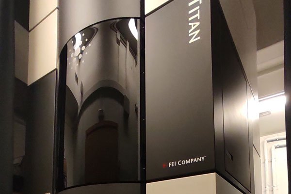
This Transmission Electron Microscope works at voltages between 60 and 300 kV. It is located in a “box” (cube) to avoid mechanical and thermal perturbations. It has a normal FEG (Shottky emitter) and a Gatan 2k x 2k CCD camera for (HR)TEM images acquisition. The main working modes in this microscope are:
- HREM: since this microscope is devoted for High Resolution Transmission Electron Microscopy (HRTEM) studies, it is equipped with a SuperTwin® objective lens and a CETCOR Cs-objective corrector from CEOS Company allowing a point to point resolution of 0.08 nm.
- STEM: In addition this Titan3 is equipped with the basic STEM facilities (BF, DF detectors) for STEM imaging at medium resolution.
- EELS: a Gatan Energy Filter Tridiem 863 allows Titan3 to perform EELS experiments in a standard and routine way (energy resolution of ̴0.7 eV).
- Lorentz and holography: as for the dedicated Titan STEM, beside these spectroscopy capabilities, the Titan3 corrected microscope is fitted with a Lorentz lens and an electrostatic biprism allowing Lorentz and medium resolution electron holography experiments to be carried on.
Image (0.08 nm resolution)
- Size and morphology information (TEM).
- Crystalline Structure (Electron Diffraction and High Resolution TEM).
- Composition: Scanning-Transmission imaging with a High Angle Annular Dark Field detector (STEM-HAADF). The contrast in the image depends on the atomic number (Z-contrast). Energy Filtered TEM (EFTEM) yields information about a specific element.
Chemical Analysis
- Electron Energy Loss Spectroscopies (EELS).
- Combined with the STEM mode: chemical composition with spatial resolution: composition maps and profiles.
Vector Field Analysis
- Electric and magnetic field studies by Electron Holography.
- Magnetic domain studies by Lorentz Microscopy.
- Stress and strain studies by HRTEM imaging.
In situ physical properties measurements
- Changes of crystalline phase (Electron diffraction)
- Defect structure by bright field/dark field imaging (BF/DF) and Weak Beam imaging (WBDF).

In Situ Formation of Carbon Nanotubes Encapsulated within Boron Nitride Nanotubes via Electron Irradiation Ref.: ACS Nano 8, 8419-8425 (2014) doi:10.1021/nn502912w

Aberration corrected HRTEM image of a magnetite nanoparticle epitaxially coated by a 1-nm-thick MgO layer. The insets show the FFT calculated from the areas marked with white squares. Ref.: Chem. Mater., 2012, 24 (3), pp 451–456. doi:10.1021/cm202306z
Laboratorio de Microscopías Avanzadas
We are a unique initiative at national and international levels. We provide the scientific and industrial community with the most advanced infrastructures in Nanofabrication, Local Probe and Electron Microscopies for the observation, characterization, nanopatterning and handling of materials at atomic and molecular scale.
Contact information
Campus Río Ebro, Edificio Edificio I+D+i

Direct Links
© 2021 LMA | Website developed by o10media | Política de privacidad | Aviso legal | Condiciones de uso | Política de Cookies |