SPIN POLARIZED STM
Performing STM scans with atomic resolution which carry simultaneously a signal proportional to the local magnetization direction, the so-called SPIN-POLARIZED STM, is available in just a few of laboratories in the world. At ELECMI-LMA, the adapted JT-STM makes this technique available to the whole scientific community. If you need to resolve the spin configuration of your surface and do not want to develop SP-STM at home from the scratch, just sent us a proposal. Some examples of the possibilities:
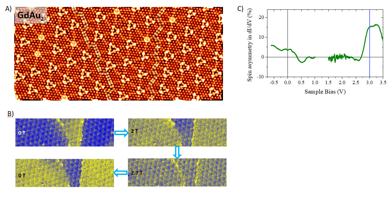
- The image in panel A is an atomically resolved topography of the Gd-Au alloy taken with a spin sensitive Cr-coated tip at 1.2 K. Panel B shows images of the differential conductance at different external magnetic fields that are taken simultaneously with the topography. The contrast in the differential conductance (color coded blue and yellow) corresponds to sample domains with opposite magnetization directions.
- The resolution of SP-STM in terms of spin contrast, is essentially the same as in STM topography one atom. This gives access to small magnetic features like the atomically sharp antiphase boundaries separating spin-up and spin-down regions in panel B.
- A typical drawback of STMs immersed in intense magnetic fields is the mechanical noise induced in the STM head. Our system passes this barrier with flying colors, with a peak-to-peak Z noise smaller than 2 pm at 0 T, remaining below 10 pm at the maximum field strength of 3 T. This provides access to the magnetic contrast evolution as a function of the magnetic field. As an example, the magnetic cycle shown in panel B drives the sample from the virgin remanent state (0 T), passing through saturation (2 T and 2.7 T), all the way back to the opposite remanent state (0 T).
- STM main difference with other microscopies is its spectroscopic capability. By taking spectra of the density of states as in regular STM spectroscopy, it is no wonder that subtracting the signals between obtained over the blue (spin-down channel) and yellow regions (spin-up channel), one gets the energy resolved spin density of the sample under study, see panel C.


Universidad de Zaragoza

Ministerio de Ciencia e Innovación

Actividad de I+D+I realizada por la Universidad de Zaragoza cofinanciada por el Gobierno de Aragón
Laboratorio de Microscopías Avanzadas
We are a unique initiative at national and international level. We provide the scientific and industrial community with the most advanced infrastructures in electron microscopy and local probe microscopy for the observation, characterization, nanostructuring and manipulation of materials at the atomic and molecular scale.
Contact information
Campus Río Ebro, Edificio Edificio I+D+i
Direct Links
© 2021 LMA | Website developed by o10media | Política de privacidad | Aviso legal | Condiciones de uso | Política de Cookies |


