

Scanning Probe Microscopies are key enabling techniques in Nanoscience and Nanotechnology, supporting a wide range of multidisciplinary activities. The LMA hosts various multipurpose AFM/STM heads that cover a broad range of applications under near ambient conditions.
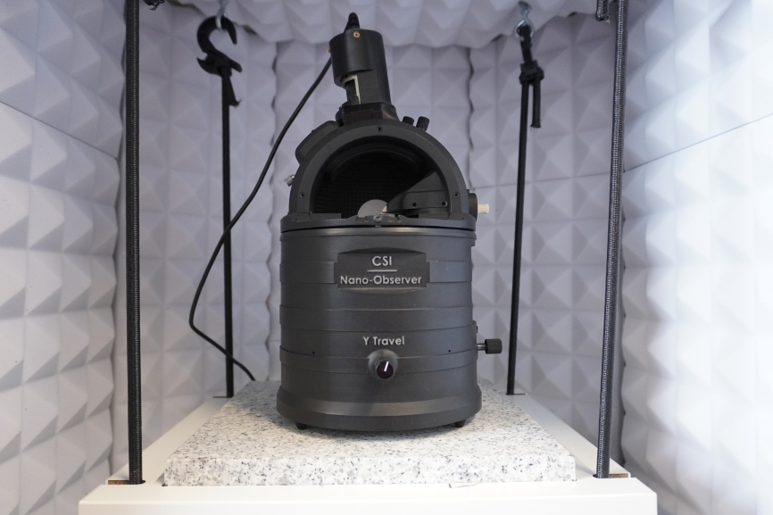
The Nano-Observer AFM from CSInstruments offers high-quality imaging in contact, intermittent contact and dynamic modes. The intermittent contact mode was developed for the scan of very soft samples where tip-sample contact is required. A specially designed Resiscope™ Preamplifier allows to measure local surface resistance over a range of 10 decades dynamically on-the-fly during conductive-AFM scans. For optically active materials like photo voltaic semiconductors the AFM’s laser can be controlled intermittently during critical measurements. All those features can be combined. The instrument is suspended in a vibration isolating cabinet allowing the operation at very low noises. A rather open design allows the sample stage to receive big samples up to 5 x 5 cm². Several AFM modes for complementary surface characterization are offered as listed below.
Key features:
- Intermittent contact mode for very soft samples;
- Resiscope™ for samples with strong variation of surface resistivity;
- Laser control for light-sensitive materials;
- Reception of big samples

- WHAT KIND OF INFORMATION CAN BE OBTAINED WITH THESE INSTRUMENTS?
- SAMPLE REQUIREMENTS
- TECHNICAL SPECIFICATIONS
- Surface morphology. Topography with resolution down to 5 Å.
- Soft Intermittent Contact
- Resiscope conductive AFM for registration for strong variation of surface resistivity
- Soft Resiscope conductive AFM for delicate samples
- Local electrical potential (KPM). Qualitative measurements of local charge distribution
- Magnetic properties (MFM). Magnetic properties analysis under magnetic fields
The sample should be immobilized onto a flat substrate (for instance, biomolecules should be immobilized onto a mica surface via adsorption or via a covalent procedure).
The sample should exhibit a lower roughness than the range of the piezo scanner.
The size of the sample should be small enough to fit inside the microscope less than 5 x 5 cm² in surface and 0.5 cm in thickness.
Types of samples that can be studied with the environmental SPMs include:
- Biological samples (DNA, proteins and peptides; cells, viruses and bacteria; biological tissues, etc.).
- Organic and inorganic thin films.
- Gels and Polymers.
- Nanoparticles and nanomaterials.
- Sensors characterization; metrology.
- Environment: Air
- Temperature Range: Room temperature
- Scanner: 100 µm(X) x 100 µm(Y) x 15 µm(Z)
Images courtesy of CSI – Concept Scientific Instruments


Laboratorio de Microscopías Avanzadas
We are a unique initiative at national and international level. We provide the scientific and industrial community with the most advanced infrastructures in electron microscopy and local probe microscopy for the observation, characterization, nanostructuring and manipulation of materials at the atomic and molecular scale.
Contact information
Campus Río Ebro, Edificio Edificio I+D+i

Direct Links
© 2023 LMA| Página web desarrollada por o10media