Different carbon and related nanomaterials, including transition metal dichalcogenides (TMD) and misfit layered compounds (MLC), are studied via different TEM techniques: high resolution (scanning) TEM imaging (HR(S)TEM), electron diffraction, electron energy loss spectroscopy (EELS), energy dispersive x-ray spectroscopy (X-EDS) and in-situ TEM approaches. It is worth mentioning that TEM studies on these beam-sensitive materials require working under particular conditions, low acceleration voltages (in our case, 60 or 80 kV) and minimizing the electron dose.
The following examples illustrate these activities:
Thermal reduction of graphene oxide (GO)
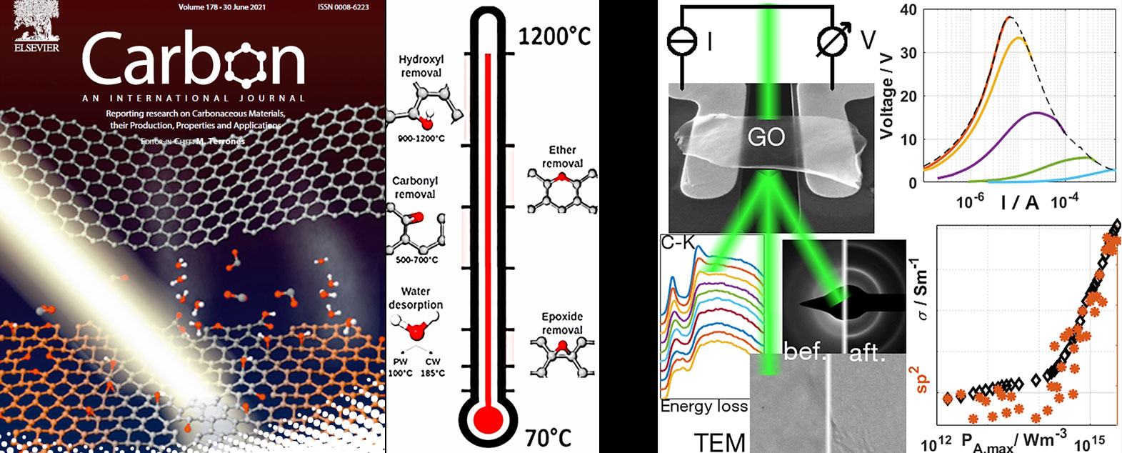
Graphene oxide (GO) strongly attracts research interest due to its use as precursor material for graphene-based material and its high flexibility in terms of functional modification offering promising applications in numerous field. Despite the efforts for investigating its atomic structure, which is critical for knowing its properties, several aspects remain unknown in particular the behavior of the physi-/chemi-sorbed and of the oxygen functional groups (OFGs) during GO reduction. Thus, we have developed two different approaches for studying, in depth, the GO thermal reduction via in-situ TEM investigations: Joule heating and heating by an external source [S. Hettler, D. Sebastian, M. Pelaez-Fdez, A. Benito, W. Maser, R. Arenal, 2D Mat. 8, 031001 (2021) ; M. Pelaez-Fernandez, A. Bermejo-Solis, A.M. Benito, W. Maser, R. Arenal, Carbon 78, 477 (2021)], see Figure 1. For achieving these comprehensive studies, we have performed HRTEM imaging, electron diffraction and EELS measurements and coupled (for this study S. Hettler, D. Sebastian, M. Pelaez-Fdez, A. Benito, W. Maser, R. Arenal, 2D Mat. 8, 031001 (2021)) with electrical conductivity measurements. In both studies we have identified the transformations of different oxygen functional groups, the desorption of physisorbed and chemisorbed water and the graphitization of the GO flakes. These in-depth analyses provide a detailed roadmap of the behavior of GO during its reduction. All these findings improve the knowledge of this complex and heterogeneous material, which is crucial for the study of their physical and chemical properties and its future applications.

Figure 1: The reduction of GO has been followed via detailed in-situ TEM (HRTEM & EELS) studies. All the processes involved in the transformation GO => r-GO have been followed for both approaches: Left & Middle: thermal [M. Pelaez-Fernandez, A. Bermejo-Solis, A.M. Benito, W. Maser, R. Arenal, Carbon 78, 477 (2021)] ; Right: Joule heating [S. Hettler, D. Sebastian, M. Pelaez-Fdez, A. Benito, W. Maser, R. Arenal, 2D Mat. 8, 031001 (2021)].
References: M. Pelaez-Fernandez, A. Bermejo-Solis, A.M. Benito, W. Maser, R. Arenal, Carbon 78, 477 (2021) and S. Hettler, D. Sebastian, M. Pelaez-Fdez, A. Benito, W. Maser, R. Arenal, 2D Mat. 8, 031001 (2021).
Functionalized carbon nanotubes
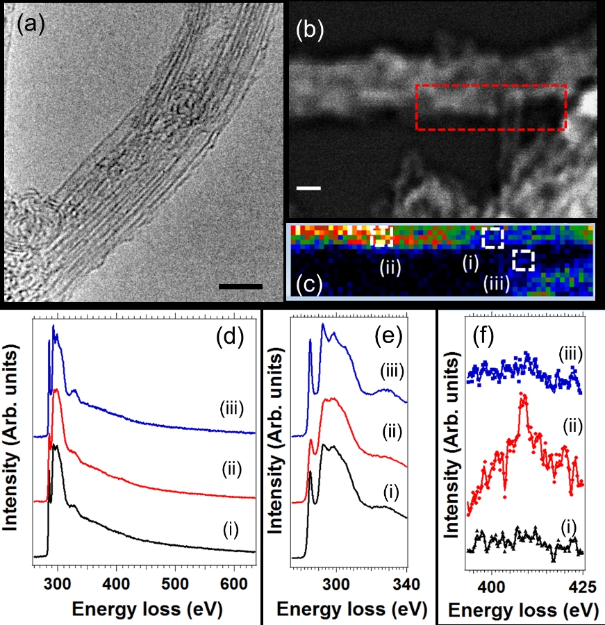
We also investigate pure and functionalized carbon nanostructures (NT, graphene, flakes, nanoparticles…). Concerning the study of the C-NTs’ functionalization via TEM, one recent example of these works is shown in Fig. 2. Here we have studied, by spatially-resolved EELS and HRTEM measurements these hybrid systems composed by C-NTs (Coll. S. Reich, Berlin Frei U. (Germany)). Thus, we have demonstrated the robustness and the stability of this system, which is very interesting as it satisfies the biocompatibility requirements.

Figure 2: (a) HRTEM micrograph of a bundle of single-walled nanotubes. (b) HAADF-STEM image of another rope of 3 or 4 single-walled CNTs. An EELS spectrum-imaging (SPIM) has been recorded in the red marked area of this image. (c) Nitrogen map extracted from the EELS-SPIM. (d) Sums of 9 selected EEL spectra collected from the three different marked areas in the SPIM, Fig. (b). While the C-K edge is visible in all the spectra, the N-K edge is only visible in the blue (Fig. (d-(ii))) EEL spectrum. (e) and (f) C- and N-K edges extracted from the EEL spectra of the Fig. (d), respectively..
Reference: A Setaro, M Adeli, M Glaeske, D Przyrembel, T Bisswanger, G Gordeev, M. Weinelt, R Arenal, R Haag, S. Reich, “Preserving p-conjugation in covalently functionalized carbon nanotubes for optoelectronic applications”, Nature Communications 8, 14281 (2017).
Misfit layered compounds (MLC) – nanotubes
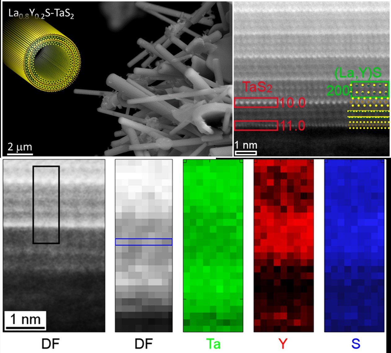
A new family of 1D nanotubes (NTs) of Misfit Layered Compounds (MLCs) has been synthesized via the chemical vapor transport technique and a comprehensive structural and compositional study of these nanostructures. In particular, high-resolution (scanning) TEM, spatially-resolved electron energy-loss spectroscopy (EELS) and electron diffraction has been developed in such NTs. These analyses demonstrate the in-phase (partial) substitution of La by Y in the (La,Y)S subsystem. The observed structure can be linked to the slightly different lattice parameters of LaS and YS as well as to an alteration of the charge transfer.

Figure caption: Atomic structure of MLC NTs. a, SEM micrograph of the tubular structures of La0.8Y0.2S-TaS2 (x = 0.2) sample and schematic illustration of such NTs. b, HAADF-STEM image of La0.8Y0.2S-TaS2 (x = 0.2) sample. The appearance of the layer stack alternates along the c-axis reveals two folding vectors rotated by 30° with respect to each other. c, EELS analysis of an NT from the same Y20 sample. Ta and Y are spatially separated between the different layers recognized in the DF image.
Reference: S. Hettler, M.B. Sreedhara, M. Serra, S.S. Sinha, R. Popovitz-Biro, I. Pinkas, A.N. Enyashin*, R. Tenne*, R. Arenal*, “YS-TaS2 and La1-xYxS-TaS2 (0≤x≤1) Nanotubes: a New Family of Misfit Layered Compounds”, ACS Nano 14, 5445–5458 (2020).
Laboratorio de Microscopías Avanzadas
We are a unique initiative at national and international levels. We provide the scientific and industrial community with the most advanced infrastructures in Nanofabrication, Local Probe and Electron Microscopies for the observation, characterization, nanopatterning and handling of materials at atomic and molecular scale.
Contact information
Campus Río Ebro, Edificio Edificio I+D+i
Direct Links
© 2021 LMA | Website developed by o10media | Política de privacidad | Aviso legal | Condiciones de uso | Política de Cookies |


